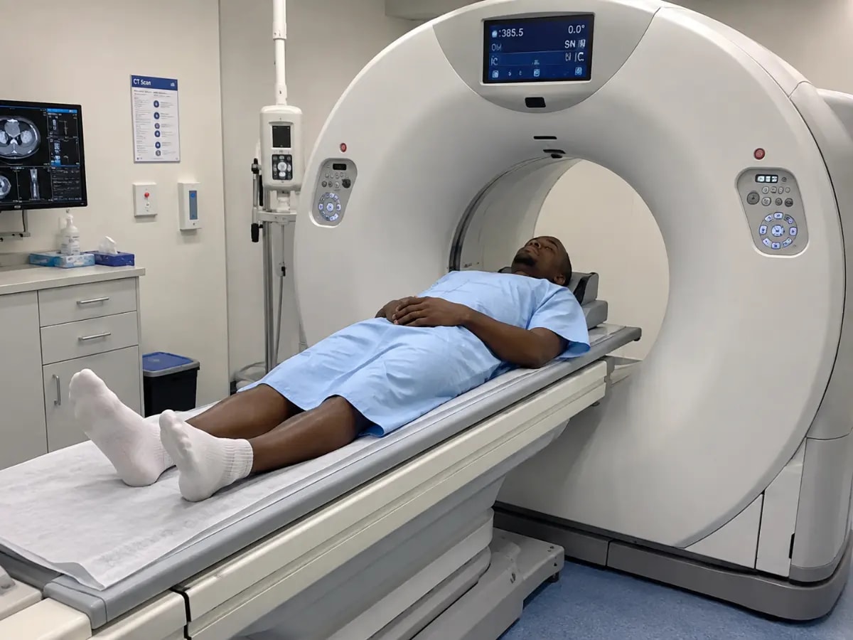
Researchers have demonstrated that tin filtered ultra low dose computed tomography (CT) produces geometric measurements of spinal pedicles comparable to those from standard dose CT. In an exploratory cadaveric study, the ultra low dose technique maintained accuracy for surgical planning of pedicle screw placement while significantly reducing radiation exposure. The findings suggest a potential shift in preoperative imaging protocols for spinal surgeries, though clinical validation is needed.
A study published in European Spine Journal has found that tin filtered ultra low dose CT scans provide morphometric data for spinal pedicles that closely aligns with measurements from standard dose CT. The research, conducted on cadaveric specimens, evaluated the geometric agreement between the two imaging techniques in the context of pedicle screw planning, a critical step in spinal stabilization procedures.
The study involved 12 cadaveric thoracic and lumbar vertebrae, each scanned using both standard dose CT and ultra low dose CT with a tin filter. Researchers measured key parameters such as pedicle width, height, and length, as well as the optimal trajectory for screw insertion. Statistical analysis revealed no clinically significant differences between the two imaging methods, with high correlation coefficients indicating strong agreement.








DISCUSSION (0)